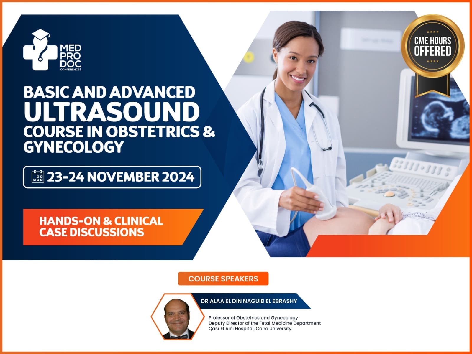
BASIC AND ADVANCED IN OBSTETRICS AND GYNECOLOGY ULTRASOUND
HANDS-ON & CLINICAL CASE DISCUSSIONS
Course Objective
Our upcoming 2023 Basic and advance in obstetrics and gynecology ultrasound course for doctors, workshops and events. It focuses on a provision in an all-inclusive understanding of Pelvic scanning including lower abdominal pain, pelvic masses. It will be in the month of Feb 4-5 organized by MED Prodoc Conference Dubai, UAE, Approved by ISUOG and Accredited by Dubai Health Authority(DHA).
Med Prodoc Conference provides ultrasound courses for doctors in dubai which covers 1st and 2nd trimester basic and advance in anatomy and physiology with advance abdominal and pelvic organs for the examination of the uterus, uterine lining (endometrium), fallopian tube and ovaries including various common pathologies relating to these organs.
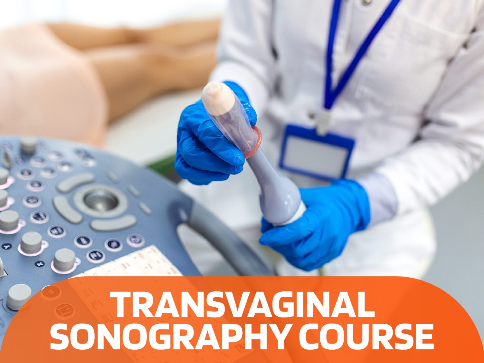
Our course includes inclusive lectures, practical sessions including obstetric gynecology ultrasound training programs, methods, transvaginal examination lectures, and practical sessions to learn fetal ultrasound. Senior and expert faculties that are a member of the ISUOG will be conducting real-time practice sessions and hands-on training for all attendees, to learn and explore about the pelvis region.
We are providing hands-on practical training workshop and is among one of the best gynecologic ultrasound training courses organization in dubai offering the world’s most modern state-of-art simulators with Accredited DHA Continuing Medical Education hours (CME) hours to upgrade knowledge and skills under internationally recognized certifications and faculties.
It is Two-days Obstetrics and gynecology ultrasound course with hands-on scanning recognized by ISUOG that focuses on a provision in an all-inclusive understanding of Pelvic scanning including lower abdominal pain, pelvic masses such as

COURSE DETAILS
Date : 2nd – 3rd March 2024
Course Type: Hands-On & Clinical Case Discussion
Minimum Qualifications: MBBS
Who Can Attend: Obstetrics and gynecologist, sonographers, Ultrasound technicians & radiologists
ISUOG Membership: Pending for Approval
SCIENTIFIC PROGRAM HIGHLIGHT
Obstetric ultrasound training programs include differential diagnosis and its method with live case study discussions with the attendees.


Explanations on procedures such as chorionic villus sampling, Placenta and amniotic fluid evalution (Amniocentesis), nuchal translucency screening and biophysical profiles.
Attendees acquire how to overcome and controls different situations and complications and the study of WHO guidelines, safety and techniques.
The ultrasound course consists of a mixture of theory and practical workshop including a demonstration that ensure proficiency for every delegate.
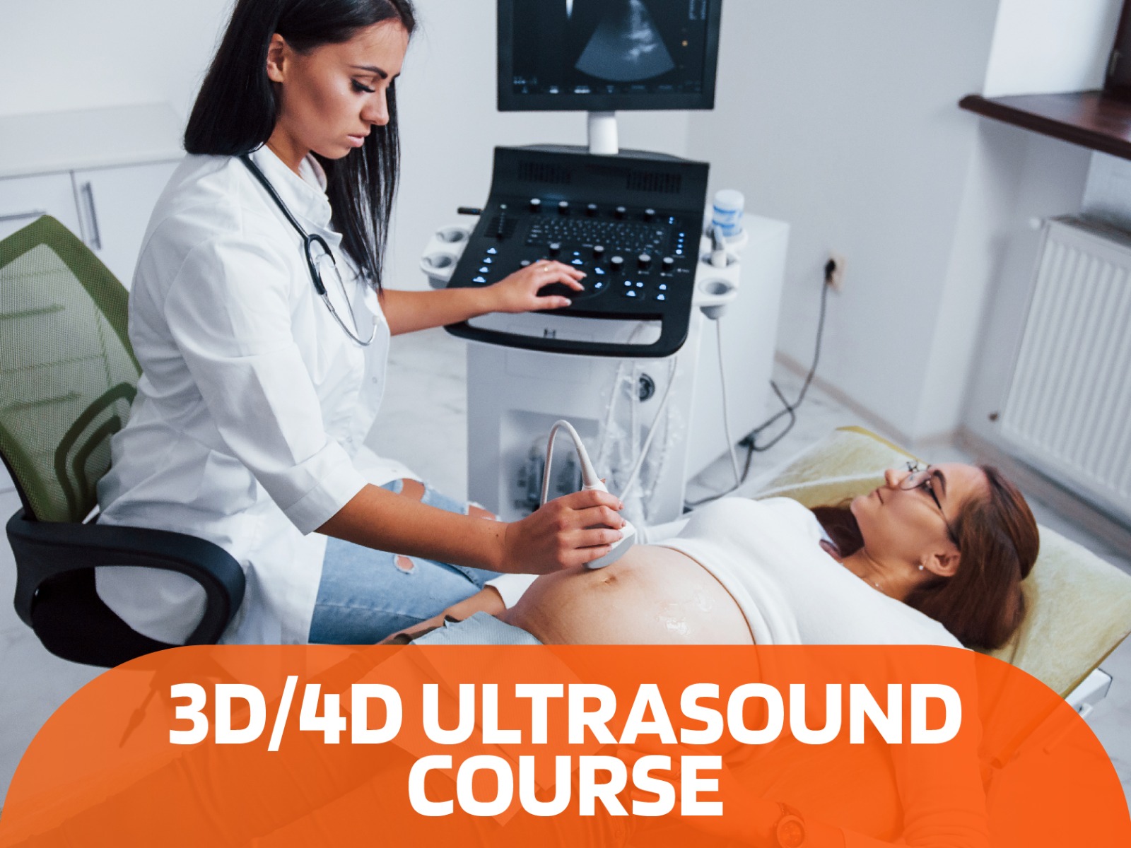
The ultrasound image is also called ultrasound scanning or sonography. Obstetric ultrasound uses sound waves to obtain pictures of embryo or fetus within a pregnant woman as well as the women uterus and ovaries and this is a perfect method to monitoring a pregnant lady and their unborn babies and the images are captured in real-time, they can show the structure and movement of the body’s internal organs. They can also show blood flowing through blood vessels.
During an obstetric ultrasound, the examiner may evaluate blood flow in the umbilical cord or may, in some cases, assess blood flow in the fetus or placenta.
Ob gyn ultrasound imaging is a noninvasive medical test that helps physicians diagnose and treat medical conditions. Ultrasound is nondestructive testing of products and structures, ultrasound is used to detect invisible flaws.
Our ultrasound training courses lectures topic “first trimester of pregnancy” which performs between 5th and 6th week able to see the gestational and yolk sacs and it allow your doctor to confirm your pregnancy. If your ultrasound is completed between weeks 6 and 7 baby which is called an embryo is measuring nearly 9 mm in length and a heartbeat is seen within this timeframe.
Ultrasounds scans during 8th to 11th weeks will show your growing baby, during this time the fetus has identifiable features such as the body, head, arms, and legs, and is moving quite readily within the gestational sac. 2
Your doctor will discuss with you in detail the need for more frequent ultrasound evaluations based on your personal situation. Additional ultrasounds may be recommended in certain cases, especially if your pregnancy is categorized as high-risk , or if you are undergoing procedures such as chorionic villus sampling, an amniocentesis, nuchal translucency screening or biophysical profiles.
The topic more dense intensive ob gyn ultrasound courses tops are “The second trimester ultrasound” which remains an important screening tool for detecting fetal abnormalities.
Medprodoc ultrasound courses for doctors include guidelines for the second trimester and ultrasound procedures designed to assist practitioners to produce high quality diagnostic survey of the fetus by demonstrating and describing recommended audio video imagining.
In medicine, ultrasound is used to detect changes in the appearance of organs, tissues, and vessels and to detect abnormal masses, such as tumors. Video clips or DVD recording of the scan has the advantage of providing moving images, which is particularly helpful when assessing the fetal heart.
COURSE SPEAKERS
Prof. Rasha Kamel
Senior Consultant at the Fetal Medicine,
Unit (CAIFM) at Cairo University
Prof. Sherif Negm
Senior Consultant at the Fetal Medicine,
Unit (CAIFM) at Cairo University
ULTRASOUND COURSES


